
INTERIORDECOR.BIZ.ID – Light microscopy, a cornerstone of scientific investigation, utilizes visible light and a system of lenses to magnify and view specimens that are too small to be seen with the naked eye. This fundamental technique allows us to explore the intricate details of cellular structures and other microscopic phenomena. It’s essential for understanding the world at a scale previously inaccessible to human observation.
Light itself is a powerful medium for interaction with the environment, influencing perception and processes across the natural world. As noted, light from the Sun not only warms the Earth and drives weather but also initiates life-sustaining biological processes. Similarly, the controlled use of light in microscopy reveals the fundamental building blocks of life.
What is Light Microscopy?
At its core, light microscopy is a method of enlarging the appearance of small objects. It works by passing light through a specimen, and then through a series of magnifying lenses. These lenses, typically made of glass, bend the light rays to create a magnified image that can be viewed by the observer or captured by a camera. The resolution and magnification achieved depend on the quality of the lenses and the wavelength of light used.
This process is analogous to how a magnifying glass works, but with much greater precision and power. The fundamental principle involves concentrating light onto a sample and then collecting and focusing the light that has interacted with the sample to form an enlarged image.
Key Components of a Light Microscope
A standard compound light microscope has several essential parts. The light source, often a bulb or LED, illuminates the specimen. The stage is where the slide containing the specimen is placed, and it can be moved to scan the sample. Above the stage, objective lenses provide initial magnification, and an eyepiece lens (or ocular lens) further magnifies the image for viewing.
Other crucial components include the diaphragm, which controls the amount of light passing through the specimen, and focusing knobs (coarse and fine) that adjust the distance between the objective lens and the specimen to achieve a sharp image. Together, these parts work in concert to produce a clear, magnified view.
How Light Microscopy Works
The process begins with the light source illuminating the specimen from below. The light passes through a condenser lens, which focuses the light onto the sample on the slide. As light rays pass through the specimen, they are affected by its different parts, either being absorbed, reflected, or refracted. These subtle changes in the light rays are what create contrast in the final image.
The magnified image then travels up through the objective lens, which performs the primary magnification. This intermediate image is then further magnified by the eyepiece lens, which the observer looks through. The final magnified image seen by the user is a virtual image, projected into their field of view.
Magnification vs. Resolution
It’s important to distinguish between magnification and resolution in microscopy. Magnification refers to how much larger the specimen appears compared to its actual size. Resolution, on the other hand, is the ability to distinguish between two closely spaced points as separate entities. High resolution is critical for seeing fine details, not just a larger blur.
For example, you can magnify an image immensely, but if its resolution is poor, you won’t be able to discern any meaningful detail. The resolution of a light microscope is limited by the wavelength of visible light, typically around 200 nanometers (0.2 micrometers).
Types of Light Microscopy
There are several variations of light microscopy, each designed to enhance contrast or visualize specific features. Bright-field microscopy is the most common, where the specimen appears darker against a bright background. Dark-field microscopy, conversely, illuminates the specimen from the sides, making it appear bright against a dark background, which is excellent for observing live, unstained organisms.
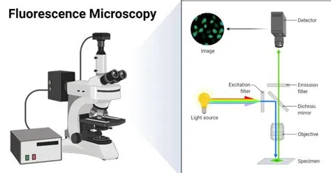
Phase-contrast microscopy is used to view unstained, transparent specimens by exploiting differences in refractive indices. Fluorescence microscopy uses fluorescent dyes to label specific structures, making them glow under UV light, revealing their location and distribution within cells. Confocal microscopy provides even higher resolution by using a pinhole aperture to eliminate out-of-focus light, creating sharp, optical cross-sections of the specimen.
Applications of Light Microscopy
Light microscopy has revolutionized numerous fields, including biology, medicine, and materials science. In biology, it’s indispensable for studying cell structure, identifying microorganisms like bacteria and fungi, and observing cellular processes such as cell division. Medical professionals use it for diagnosing diseases by examining blood, tissue samples, and identifying pathogens.
Materials scientists use light microscopes to examine the structure and defects in materials like metals, polymers, and ceramics. Even in forensic science, light microscopy aids in the analysis of evidence like fibers, hair, and paint chips. The versatility of light microscopy makes it a foundational tool for countless scientific discoveries and practical applications.
The Importance of Light
The very essence of light microscopy is the intelligent application of light. Just as natural light enables organisms to perceive and interact with their surroundings, artificial light in a microscope allows us to perceive and understand the microscopic world. This controlled illumination reveals structures and dynamics that would otherwise remain hidden, driving scientific progress.
The ability to manipulate and observe how light interacts with matter at this scale is what makes microscopy such a powerful scientific endeavor. From the fundamental building blocks of life to the structural integrity of advanced materials, light microscopy provides the window through which we can see and understand.
Limitations and Future Directions
Despite its power, light microscopy has inherent limitations, primarily related to resolution, which is dictated by the wavelength of light. To overcome these limits for viewing even smaller structures like viruses or individual molecules, scientists turn to electron microscopy. However, light microscopy remains the most accessible and widely used technique for routine biological and materials analysis.
Ongoing advancements in light microscopy, such as super-resolution techniques, are pushing the boundaries of what can be seen, offering even greater detail. These innovations continue to expand the capabilities of this foundational scientific tool, ensuring its relevance for years to come.
Frequently Asked Questions (FAQ)
What is the primary function of light in microscopy?
The primary function of light in microscopy is to illuminate the specimen and then be transmitted, reflected, or diffracted by it. This interaction with the specimen alters the light in a way that, when magnified by lenses, creates a visible image revealing the specimen’s structures.
Can you see individual atoms with a light microscope?
No, individual atoms cannot be seen with a standard light microscope. The resolution of light microscopy is limited to about 200 nanometers, which is far too large to resolve atomic structures. Electron microscopy is required for imaging at the atomic scale.
What is the difference between magnification and resolution?
Magnification is the process of enlarging the apparent size of an object, making it look bigger. Resolution, however, is the ability to distinguish between two closely spaced objects as separate entities. High resolution is crucial for seeing fine details, whereas high magnification without good resolution results in a blurry, indistinct image.
Why is light microscopy still important today?
Light microscopy remains important due to its relative affordability, ease of use, and versatility. It is ideal for viewing live cells and dynamic processes, observing larger cellular structures, and performing routine diagnostics in medicine and research where extreme resolution is not always necessary.
Written by: Emma Johnson