
INTERIORDECOR.BIZ.ID – Light is a fundamental element in our interaction with the world, powering everything from our perception to the very existence of life on Earth. This intrinsic connection to light extends to our scientific endeavors, particularly in understanding the microscopic realm.
When we discuss the ‘light definition microscope,’ we are referring to instruments that utilize light as their primary source for illumination and magnification. These are the foundational tools that revolutionized our ability to see beyond the limitations of the naked eye.
The Essence of Light in Microscopy
The principle behind a light definition microscope is straightforward: light passes through or reflects off a specimen, and then through a series of lenses. These lenses magnify the image, allowing us to observe details invisible to us otherwise.
Without light, the complex optical systems within a microscope would have nothing to transmit or reflect, rendering them useless for viewing. The quality and properties of the light directly impact the clarity and resolution of the image observed.
How Light Enables Magnification
A light microscope works by bending light rays through lenses. The specimen is placed on a stage, and a light source, typically located below the stage, illuminates it. The transmitted light then travels upwards through the objective lens and the eyepiece lens.
Each lens system in the microscope contributes to the overall magnification. The objective lens provides the initial, higher magnification, and the eyepiece lens further magnifies this intermediate image for the observer’s eye.
Types of Light Microscopes
The most common type is the bright-field microscope, where the specimen appears dark against a bright background. This is the workhorse for many basic biological and material science observations.
Other variations exist, such as dark-field microscopes, which illuminate specimens from the sides, making them appear bright against a dark background, enhancing visibility of unstained samples.
Phase Contrast and Fluorescence Microscopy
Phase contrast microscopy is particularly useful for viewing living, unstained cells. It converts differences in light refraction and density within the specimen into variations in brightness, revealing internal structures.
Fluorescence microscopy uses specific wavelengths of light to excite fluorescent molecules (fluorophores) within the specimen. These molecules then emit light at longer wavelengths, creating a brightly colored image against a dark background, allowing for highly specific targeting of cellular components.
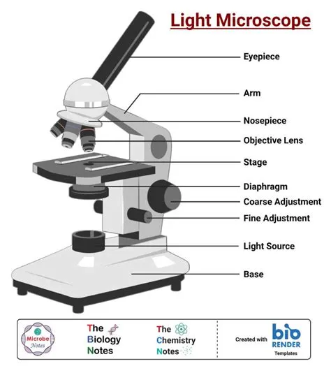
Key Components of a Light Microscope
A typical light microscope comprises several essential parts. The light source, often an LED or halogen lamp, provides the illumination necessary for viewing. The diaphragm controls the amount of light passing through the specimen, affecting contrast and brightness.
The stage is where the specimen slide is placed, usually with clips to hold it in position. Coarse and fine adjustment knobs are crucial for focusing the image, bringing the specimen into sharp view.
Lenses: The Heart of Magnification
The objective lenses, mounted on a revolving nosepiece, offer different levels of magnification (e.g., 4x, 10x, 40x, 100x). The higher the magnification of the objective, the closer you can get to observing fine details.
The eyepiece lens, or ocular lens, is where the user looks. It further magnifies the image produced by the objective lens, typically by 10x. The total magnification is the product of the objective lens magnification and the eyepiece lens magnification.
The Impact of Light Definition Microscopes
The invention and refinement of the light definition microscope have been pivotal in countless scientific discoveries. From identifying bacteria and understanding cell structures to diagnosing diseases, its impact is immeasurable.
These instruments democratized the study of the microscopic world, making complex biological and chemical processes observable and understandable for researchers and students alike.
Illuminating the Invisible
The concept of using light to see the unseen is the core principle that drives these powerful tools. The ability to manipulate light, enhance its properties, and precisely focus it has allowed humanity to explore realms previously hidden from view.
Understanding how a light definition microscope works empowers users to optimize their observations, select the appropriate microscopy techniques, and interpret the images they acquire with greater accuracy.
FAQ Section
What is the primary function of light in a microscope?
Light in a microscope serves as the illumination source that passes through or reflects off a specimen, enabling it to be magnified and viewed.
How does a light microscope achieve magnification?
Magnification is achieved through a system of lenses, specifically objective lenses and eyepiece lenses, which bend and enlarge the image of the specimen.
What is the difference between bright-field and dark-field microscopy?
Bright-field microscopy shows specimens as dark against a bright background, while dark-field microscopy illuminates specimens brightly against a dark background.
Why is phase contrast microscopy useful for living cells?
Phase contrast microscopy allows visualization of unstained living cells by converting differences in light refraction into visible contrast.
What are fluorophores used for in microscopy?
Fluorophores are used in fluorescence microscopy to make specific cellular components or molecules visible by emitting light after being excited by specific wavelengths.
Frequently Asked Questions (FAQ)
What is the primary function of light in a microscope?
Light in a microscope serves as the illumination source that passes through or reflects off a specimen, enabling it to be magnified and viewed.
How does a light microscope achieve magnification?
Magnification is achieved through a system of lenses, specifically objective lenses and eyepiece lenses, which bend and enlarge the image of the specimen.
What is the difference between bright-field and dark-field microscopy?
Bright-field microscopy shows specimens as dark against a bright background, while dark-field microscopy illuminates specimens brightly against a dark background.
Why is phase contrast microscopy useful for living cells?
Phase contrast microscopy allows visualization of unstained living cells by converting differences in light refraction into visible contrast.
What are fluorophores used for in microscopy?
Fluorophores are used in fluorescence microscopy to make specific cellular components or molecules visible by emitting light after being excited by specific wavelengths.
Written by: David Thomas