
INTERIORDECOR.BIZ.ID – Light is a fundamental force, instrumental in how many organisms perceive and interact with their environment. The sun’s illumination, for instance, provides warmth, influences weather, and is the initial spark for life-sustaining processes on Earth.
In biology, understanding the intricate structures of life often requires magnification beyond what the human eye can achieve. This is where the light microscope definition biology comes into play, offering a crucial window into the microscopic world.
What is a Light Microscope?
A light microscope, also known as an optical microscope, is a scientific instrument that uses visible light and a system of lenses to magnify small objects. It allows us to see details that are too small to be observed with the naked eye, revealing the complex architecture of cells, tissues, and microorganisms.
These microscopes are ubiquitous in biological research and education, forming the bedrock of many investigations into living organisms. Their accessibility and relative simplicity make them an indispensable tool for exploring the foundational units of life.
How Does a Light Microscope Work?
The basic principle involves passing light through a specimen and then through a series of magnifying lenses. The light source, typically a lamp or mirror, illuminates the sample placed on a stage.
The specimen is usually mounted on a glass slide and covered with a coverslip. The magnified image is then viewed through one or more eyepieces, known as ocular lenses, and sometimes also through objective lenses.
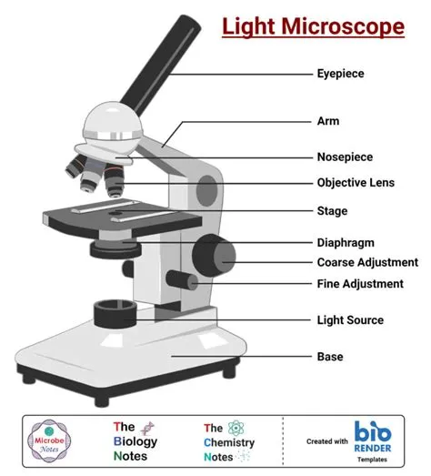
Key Components of a Light Microscope
Several essential parts work in concert to produce a magnified image. The illumination system provides the light, while the stage holds the specimen for viewing.
The objective lenses, located near the specimen, provide the initial magnification, and the eyepiece lenses further enlarge this image for the observer. Focusing knobs allow for fine adjustments to ensure a clear image.
Types of Light Microscopy
Various techniques enhance the capabilities of light microscopy. Bright-field microscopy, the most common type, involves light passing directly through the specimen, creating a darkened image against a bright background.
Phase-contrast microscopy is used for viewing unstained, transparent specimens by exploiting differences in light refraction. This technique is particularly useful for observing live cells without the need for staining, which can sometimes alter cell behavior.
Other Important Techniques
Dark-field microscopy, in contrast, illuminates the specimen from the sides, making it appear bright against a dark background, which is excellent for observing fine details of thin structures.
Fluorescence microscopy utilizes fluorescent dyes that emit light when excited by specific wavelengths, allowing for the visualization of specific molecules or structures within cells.
Applications in Biology
The light microscope definition biology is central to countless biological disciplines. It is used to study cell structure, identify bacteria and other microorganisms, and analyze tissue samples in histology.
Researchers use light microscopes to observe cellular processes like cell division and movement, and to diagnose diseases by examining blood cells, parasites, and other biological samples.
Exploring the Microscopic World
From understanding the single-celled organisms that form the base of many ecosystems to examining the intricate organization of multicellular tissues, the light microscope is an indispensable tool.
It demystifies the unseen world, revealing the beauty and complexity of life at its most fundamental levels and driving scientific discovery forward.
Limitations of Light Microscopy
Despite its versatility, light microscopy has resolution limits. The resolving power of a microscope is its ability to distinguish between two closely spaced points, and for light microscopes, this is typically around 200 nanometers.
This means that structures smaller than this, such as viruses or the detailed internal components of organelles, cannot be resolved with standard light microscopy and require more advanced techniques like electron microscopy.
The Importance of Magnification and Resolution
Magnification refers to how much larger the image appears compared to the actual object, while resolution is about the clarity and detail of that magnified image. High magnification without good resolution results in a blurry, uninformative image.
Light microscopes achieve magnification through their lens systems, but their resolution is fundamentally limited by the wavelength of visible light, a key factor in its biological applications and limitations.
Conclusion
In conclusion, the light microscope definition biology highlights its role as a cornerstone instrument. It enables us to visualize and study the microscopic components of life, from single cells to complex tissues, laying the groundwork for advancements in medicine, agriculture, and environmental science.
Its continued use and the development of advanced light microscopy techniques underscore its enduring significance in biological research and education, making the unseen world accessible and understandable.
Frequently Asked Questions about Light Microscopes
Question: What is the primary function of a light microscope in biology?
Answer: The primary function of a light microscope in biology is to magnify small objects and structures that are invisible to the naked eye, allowing scientists to study cells, tissues, and microorganisms in detail.
Question: What is the difference between magnification and resolution in microscopy?
Answer: Magnification refers to the enlargement of an object’s image, while resolution is the ability to distinguish between two closely spaced objects. A good microscope needs both high magnification and high resolution to provide clear and detailed images.
Question: Can light microscopes view viruses?
Answer: Standard light microscopes typically cannot resolve viruses because viruses are smaller than the resolving power of light microscopes, which is around 200 nanometers. Electron microscopes are needed to visualize viruses.
Question: What are some common types of light microscopy used in biology?
Answer: Common types include bright-field microscopy, phase-contrast microscopy, dark-field microscopy, and fluorescence microscopy, each offering different ways to visualize specimens based on their optical properties or the use of fluorescent labels.
Question: Why are light microscopes important for studying live cells?
Answer: Techniques like phase-contrast microscopy allow for the observation of live cells without staining, which can kill or alter the cells. This enables the study of dynamic cellular processes in their natural state.
Written by: William Clark